Human Anatomy Rib Cage Muscles : Internal Intercostal An Overview Sciencedirect Topics / It encloses the thoracic cavity, which contains the lungs.. The human rib cage is made up of 12 paired rib bones; The middle and upper part of your spine is called the thoracic region and it helps to support your upper body. Its functions are to protect the thoracic organs from trauma and also form the bony attachment for various muscles. An inhalation is accomplished when the muscular diaphragm, at the floor of the thoracic cavity, contracts and flattens, while the contraction of intercostal muscles lift the rib cage up and out. The transversus thoracic muscles originate from the posterior surface of the xiphoid process and the lower part of the body of the sternum.
It encloses and protects the heart and lungs. The fibres pass superolaterally to insert into the costal cartilages of ribs three to six. The upper edge is round and the lower sharp. In this image, you will find thoracic vertebrum, costochondral joint, costal cartilage, costal margin, costal arch, thoracic vertebrum, xiphoid process, xiphisternal joint, body, manubrial sternal joint, manubrium, the sternal notch in it. An inhalation is accomplished when the muscular diaphragm, at the floor of the thoracic cavity, contracts and flattens, while the contraction of intercostal muscles lift the rib cage up and out.
Lawyersnaperville Org All About Human Anatomy Human Body Anatomy Human Body Diagram Human Body Systems from i.pinimg.com Rib cage, in vertebrate anatomy, basketlike skeletal structure that forms the chest, or thorax, and is made up of the ribs and their corresponding attachments to the sternum (breastbone) and the vertebral column. The primary job of flat bones is to protect underlying structures. The area under the ribs consists of intercostal muscle, ligaments and tendons, as well as the abdominal obliques, transverus abdominis and rectus abdominis just below the rib cage. The ribs are part of the axial skeleton and are classified as flat bones. Diagram of human body, liver rib cage, rib cage diagram labeled, rib cage diagram numbered, rib cage diaphragm, rib cage heart, rib cage organs anatomy, rib cage pain, stomach, diagram of human body, liver rib cage, rib cage diagram labeled, rib cage diagram numbered, rib cage diaphragm, rib cage. Anatomy of the rib cage diagram. A rib has a flat body, as you can see from the picture of the anatomy of the human rib cage. Related posts of muscle anatomy rib cage muscle.
An inhalation is accomplished when the muscular diaphragm, at the floor of the thoracic cavity, contracts and flattens, while the contraction of intercostal muscles lift the rib cage up and out.
Its functions are to protect the thoracic organs from trauma and also form the bony attachment for various muscles. At the chest, many rib bones connect to the sternum via costal cartilage,. Other flat bones in the human body are found in the pelvis and skull. Related posts of muscle anatomy rib cage muscle. In this image, you will find thoracic vertebrum, costochondral joint, costal cartilage, costal margin, costal arch, thoracic vertebrum, xiphoid process, xiphisternal joint, body, manubrial sternal joint, manubrium, the sternal notch in it. It has clear front side and back planes. An inhalation is accomplished when the muscular diaphragm, at the floor of the thoracic cavity, contracts and flattens, while the contraction of intercostal muscles lift the rib cage up and out. The rib cage is often simplified as an oval shape. A rib has a flat body, as you can see from the picture of the anatomy of the human rib cage. In this video, we explore:1) the anatomy of the sternum2) the anatomy and differences between the three classes of ribs3) the anatomy and differences between. Browse 6,946 human rib cage stock photos and images available, or search for human heart or skeleton to find more great stock photos and pictures. Über 7 millionen englischsprachige bücher. The human rib cage (thoracic cage) has the very important job of protecting the heart and lungs.
Anatomy of the rib cage diagram. Rib cage, in vertebrate anatomy, basketlike skeletal structure that forms the chest, or thorax, and is made up of the ribs and their corresponding attachments to the sternum (breastbone) and the vertebral column. In this video, we explore:1) the anatomy of the sternum2) the anatomy and differences between the three classes of ribs3) the anatomy and differences between. It has clear front side and back planes. Human anatomy drawing human figure drawing anatomy study anatomy art anatomy reference figure drawing reference pose reference anatomy bones body anatomy.
Thoracic Wall And Breast Illustrations from www.imaios.com The ribs are part of the axial skeleton and are classified as flat bones. Browse 6,946 human rib cage stock photos and images available, or search for human heart or skeleton to find more great stock photos and pictures. It may occur after an obvious injury or without explanation. The rib cage has three important functions: The human rib cage (thoracic cage) has the very important job of protecting the heart and lungs. It is also the center around which the superior 10 ribs directly or indirectly attached. With the upper ribs, closer to the nodule (and in the case of lower ribs, a little further from the nodule) they are curved and have a rough surface that connects them with muscles, angulus costae. In this video, we explore:1) the anatomy of the sternum2) the anatomy and differences between the three classes of ribs3) the anatomy and differences between.
With the upper ribs, closer to the nodule (and in the case of lower ribs, a little further from the nodule) they are curved and have a rough surface that connects them with muscles, angulus costae.
Browse 6,946 human rib cage stock photos and images available, or search for human heart or skeleton to find more great stock photos and pictures. Human muscles · april 17, 2020. The human rib cage (thoracic cage) has the very important job of protecting the heart and lungs. The ribs are part of the axial skeleton and are classified as flat bones. It may occur after an obvious injury or without explanation. Jun 17, 2021 · anterior shoulder muscles, also called the pectoral muscles, attach the upper extremity to the clavicle and the thoracic cage. #proko #art #anatomy #ribs #ribcage #humananatomy #tutorial. The rib cage is often simplified as an oval shape. A rib has a flat body, as you can see from the picture of the anatomy of the human rib cage. The area under the ribs consists of intercostal muscle, ligaments and tendons, as well as the abdominal obliques, transverus abdominis and rectus abdominis just below the rib cage. It encloses the thoracic cavity, which contains the lungs. In this video, we explore:1) the anatomy of the sternum2) the anatomy and differences between the three classes of ribs3) the anatomy and differences between. Rib cage pain can be caused.
In this image, you will find thoracic vertebrum, costochondral joint, costal cartilage, costal margin, costal arch, thoracic vertebrum, xiphoid process, xiphisternal joint, body, manubrial sternal joint, manubrium, the sternal notch in it. It encloses and protects the heart and lungs. The upper edge is round and the lower sharp. This muscle assists in depression of the ribs. Related posts of muscle anatomy rib cage muscle.
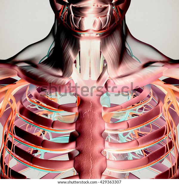
Human Anatomy Torso Rib Cage Muscle Stock Illustration 429363307 from image.shutterstock.com Muscles of the spine and 8 rib muscles anatomy rib muscles anatomy and human anatomy muscles rib cage diagram. Human anatomy drawing human figure drawing anatomy study anatomy art anatomy reference figure drawing reference pose reference anatomy bones body anatomy. It is also the center around which the superior 10 ribs directly or indirectly attached. Other flat bones in the human body are found in the pelvis and skull. The human rib cage is a component of the human respiratory system. It provides a strong framework onto which the muscles of the shoulder girdle, chest, upper abdomen and back can attach. The rib cage has three important functions: At the chest, many rib bones connect to the sternum via costal cartilage,.
The human rib cage is made up of 12 paired rib bones;
Rib cage pain can be caused. Rib cage pain may be sharp, dull, or achy and felt at or below the chest or above the navel on either side. The ribs are part of the axial skeleton and are classified as flat bones. It encloses the thoracic cavity, which contains the lungs. The human rib cage (thoracic cage) has the very important job of protecting the heart and lungs. Other flat bones in the human body are found in the pelvis and skull. The middle and upper part of your spine is called the thoracic region and it helps to support your upper body. An inhalation is accomplished when the muscular diaphragm, at the floor of the thoracic cavity, contracts and flattens, while the contraction of intercostal muscles lift the rib cage up and out. The transversus thoracic muscles originate from the posterior surface of the xiphoid process and the lower part of the body of the sternum. In this image, you will find thoracic vertebrum, costochondral joint, costal cartilage, costal margin, costal arch, thoracic vertebrum, xiphoid process, xiphisternal joint, body, manubrial sternal joint, manubrium, the sternal notch in it. This muscle assists in depression of the ribs. It encloses and protects the heart and lungs. Our latest youtube film is ready to run.


0 Komentar